

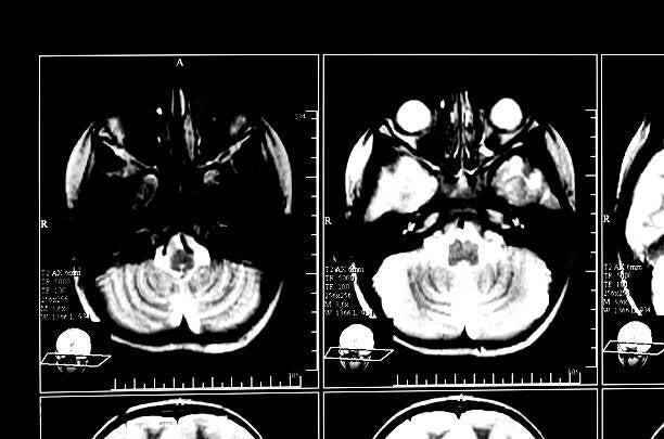
A resident scrolling through a TMJ MRI for the first time often faces the same question: which side is the disc actually on? At 3D Maxillofacial Diagnostics, this is one of the most common teaching moments in oral radiology, because disc displacement is common, frequently missed on a quick read, and genuinely confusing until you understand exactly what you are looking for on the open and closed mouth MRI TMJ series.
This guide walks through what does disc displacement look like on MRI, how open and closed mouth TMJ MRI sequences work together to establish a diagnosis, and how to reliably tell disc displacement with reduction apart from disc displacement without reduction, the single most important distinction in TMJ MRI interpretation.
Why MRI Is the Gold Standard for TMJ Disc Evaluation
Unlike CBCT, which shows bone in exceptional detail but cannot visualize the articular disc, MRI is the only imaging modality that directly demonstrates the soft tissue anatomy of the temporomandibular joint. The disc itself, a biconcave fibrocartilaginous structure sitting between the condylar head and the articular eminence, is essentially invisible on any bone-based imaging. If a patient has clicking, locking, or limited opening and the clinical picture points toward an internal derangement, MRI is the modality that actually answers the question.
This is exactly why TMJ MRI interpretation is treated as its own diagnostic skill set rather than an extension of general radiographic reading. The disc is small, it moves, and its position has to be assessed relative to the condyle in two different jaw positions before a diagnosis can be made with confidence.
The Basic Anatomy Every Reader Should Know
Before interpreting a scan, it helps to fix three landmarks in your mind:
- The condylar head, the rounded bony structure that the disc sits on top of
- The disc, seen as a biconcave dark (low signal) structure on T1 and proton-density sequences, thicker at its anterior and posterior bands and thinner centrally
- The posterior attachment or bilaminar zone, the vascularized soft tissue behind the disc that stretches as the disc displaces forward
In a normal joint, the posterior band of the disc sits at the twelve o'clock position directly above the condylar head in the closed-mouth view. This reference point, often described using a clock-face or a line drawn perpendicular to the long axis of the condyle, is the anchor for every disc position assessment that follows.
Open and Closed Mouth MRI TMJ: Why Both Positions Matter
A single closed-mouth image only tells half the story. The open and closed mouth MRI TMJ protocol is what allows a reader to distinguish a disc that is displaced but still functional from one that is displaced and mechanically blocking normal movement. The standard protocol includes:
- Closed-mouth sagittal images (T1 and proton-density or T2), used to establish baseline disc position relative to the condyle
- Open-mouth sagittal images, captured with the mouth open to maximum comfortable opening, used to see whether the disc recaptures a normal position over the condyle as it translates forward
- Coronal images in at least one jaw position, used to assess for medial or lateral disc displacement, which sagittal images alone can miss
- T2-weighted sequences, useful for identifying joint effusion, a fluid signal that often correlates with active inflammation or a painful joint
Skipping the open-mouth series is one of the most common reasons a disc displacement gets mischaracterized. A disc that looks displaced on the closed-mouth image alone could reduce completely on opening, which changes both the diagnosis and the management plan.
What Does Disc Displacement Look Like on MRI?
In practical terms, disc displacement means the disc's posterior band has moved away from the twelve o'clock reference position, almost always anteriorly, sometimes with a medial or lateral component. On the closed-mouth image, instead of sitting directly above the condyle, the posterior band appears anterior to it, and the thinner intermediate zone of the disc may be sitting where the posterior band should be. The bilaminar zone posterior to the disc often appears stretched or thinned as it is pulled anteriorly along with the disc.
Once displacement is identified on the closed-mouth series, the open-mouth image determines what kind of displacement it is:
- If the disc returns to a normal position over the condylar head as the mouth opens, this is disc displacement with reduction
- If the disc remains anteriorly displaced throughout opening and never recaptures a normal position, this is disc displacement without reduction
This single comparison, closed-mouth position versus open-mouth position, is the crux of TMJ MRI interpretation and the detail residents most often rush past.
Disc Displacement With Reduction vs Without Reduction
Feature
Disc Displacement WITH Reduction
Disc Displacement WITHOUT Reduction
Closed-mouth disc position
Anteriorly (or anteromedially) displaced relative to the condylar head
Anteriorly displaced relative to the condylar head
Open-mouth disc position
Disc recaptures — returns to a normal position atop the condyle
Disc remains displaced; does not recapture
Clinical sign
Reciprocal clicking (click on opening, click on closing)
Often no click; limited mouth opening (closed lock)
Condylar translation
Typically normal or near-normal
Often restricted, condyle may not translate fully
Bilaminar zone
Usually preserved, may show mild stretching
Often shows degenerative signal change or thinning over time
Common progression
May remain stable or progress to without reduction over time
Associated more often with chronic pain and osteoarthritic change
Clinically, disc displacement with reduction is often the imaging correlate of reciprocal clicking, a click on opening as the disc recaptures, and a second click on closing as it displaces again. Disc displacement without reduction is the imaging correlate of a closed lock, restricted opening because the displaced disc is physically blocking normal condylar translation. Recognizing which pattern is present directly shapes whether a patient is managed conservatively, referred for physical therapy, or considered for more advanced intervention.
A Step-by-Step Approach to Reading a TMJ MRI
- Confirm image quality and that both open and closed mouth sequences are present and properly labeled left and right.
- Assess the closed-mouth sagittal images first, locating the posterior band of the disc relative to the twelve o'clock position on the condyle.
- Assess the open-mouth sagittal images, determining whether the disc recaptures a normal position over the condyle.
- Review coronal images to rule out a purely medial or lateral displacement, which can be missed on sagittal images alone.
- Check T2-weighted sequences for joint effusion or other signal changes suggesting active inflammation.
- Evaluate the condylar bone itself for flattening, erosion, or osteophyte formation, since chronic disc displacement often coexists with degenerative joint change.
- Correlate every imaging finding with the clinical history, since a click on exam and a click on imaging do not always mean the same thing.
Common Pitfalls in TMJ MRI Interpretation
A few mistakes come up repeatedly, even among experienced general practitioners reading their own scans:
- Reading only the closed-mouth series and assuming reduction status without the open-mouth comparison
- Missing a partial or medial-only displacement because coronal images were not reviewed
- Confusing a thinned, deformed disc (common in chronic without-reduction cases) with a normally shaped but simply displaced disc
- Overlooking joint effusion on T2 sequences, which can be an important marker of active symptoms even when disc position looks stable
- Failing to correlate bone changes on the same MRI with the disc findings, missing early osteoarthritic change
Why Educator-Level Expertise Matters Here
TMJ MRI is one of the areas where reading experience and teaching experience genuinely overlap. Dr. Shereen Shokry, the Canadian Certified Oral and Maxillofacial Radiologist behind every report at 3D Maxillofacial Diagnostics, has spent more than twenty years both interpreting these studies and teaching dental professionals and residents how to read them. That educator background matters in a TMJ imaging analysis report, because a report written by someone who also teaches this material tends to explain the finding, not just label it, which is exactly what a referring dentist needs when deciding how to counsel a patient or when to refer for further management.
This is also why residents and general dentists building their own diagnostic skills often use these reports as a learning tool alongside patient care, comparing their own read of the open and closed mouth series against a specialist interpretation.
What's Included in a TMJ MRI Report
A complete, clinically useful TMJ MRI report should cover:
- Disc position on closed-mouth images, described relative to the condylar head
- Disc position and recapture status on open-mouth images, with a clear statement of reduction versus non-reduction
- Assessment for medial or lateral displacement on coronal images
- Presence or absence of joint effusion on T2-weighted sequences
- Condylar bone assessment, including any degenerative or osteoarthritic change
- A clear summary impression correlating the imaging with the referring clinician's specific question
If you have a case that needs this level of detail, you can review the TMJ MRI report submission requirements before sending a study over.
How the Reporting Process Works
Submitting a TMJ MRI case for specialist interpretation follows a simple sequence:
- Capture or obtain both open-mouth and closed-mouth sagittal sequences, plus at least one coronal series, ideally with a T2-weighted sequence included.
- Export the original DICOM files along with any relevant clinical notes, such as click history, locking episodes, or pain patterns.
- Submit the case for review.
- Receive a structured written report explaining disc position, reduction status, and any associated joint changes, along with a clear clinical impression.
The full imaging submission process outlines exactly what file formats and clinical details are needed before a case can be reviewed.
Read the Joint With Confidence
TMJ MRI interpretation rewards a systematic approach: establish disc position on closed-mouth images, confirm reduction status on open-mouth images, check coronal images for medial or lateral displacement, and correlate every finding with the clinical picture. For complex or borderline cases, a specialist read backed by genuine teaching experience gives both the referring clinician and the patient a clear, well-explained answer rather than a label on a report.
If you have a TMJ case you would like reviewed, or questions about submission requirements, reach out directly and the team can walk you through next steps.
Frequently Asked Questions