

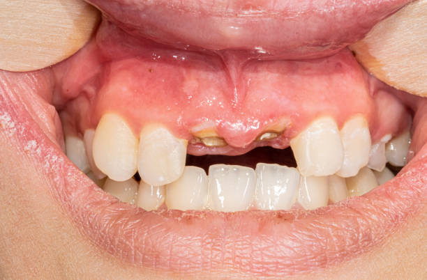
A patient returns eighteen months after root canal treatment with a dull ache, a sinus tract, or a periapical radiolucency that simply will not resolve. Is this a failed root canal that needs retreatment, an apicoectomy, or an extraction because the root itself has cracked? On a periapical film, both problems can look almost identical.
At 3D Maxillofacial Diagnostics, this is exactly the kind of case where 3D dental imaging changes the diagnostic conversation, and why more clinicians are turning to dedicated cbct interpretation services before deciding on a treatment plan.
This article walks through how do you diagnose a cracked tooth root, what distinguishes root fracture vs failed RCT on imaging, and why a trained eye reading a 3d cbct scan can save patients from an unnecessary retreatment or, just as importantly, from unnecessary tooth loss.
Why 2D Radiographs Fall Short
Periapical and panoramic radiographs remain useful first-line tools, but they compress a three-dimensional root into a two-dimensional shadow. A vertical root fracture that runs buccolingually can be completely invisible on a straight-on periapical view because the fracture line lies parallel to the beam. Overlapping anatomy, angulation errors, and the classic "halo" or "J-shaped" radiolucency associated with fractures can also mimic ordinary periapical inflammatory lesions from a failed root canal.
This is the core clinical problem: two very different diagnoses, a root fracture requiring extraction versus a failed root canal that may be retreatable, can present with strikingly similar 2D findings. Guessing wrong means either subjecting a patient to a retreatment that was never going to succeed, or extracting a tooth that could have been saved. That is why oral radiology consultation with cross-sectional imaging has become a standard of care for ambiguous endodontic failures.
What a CBCT 3D Dental Scan Actually Shows
A cbct 3d dental scan captures a volumetric dataset of the tooth and surrounding bone, which can then be sliced in axial, coronal, and sagittal planes at sub-millimeter resolution. Instead of one flattened image, the interpreting radiologist can scroll through the entire root, slice by slice, looking for:
- A discrete radiolucent line running along the length of the root, often widening apically or coronally
- A characteristic "halo" or doughnut-shaped area of bone loss surrounding the root, distinct from the typical rounded periapical lesion of endodontic origin
- Separation or displacement of root fragments
- Localized loss of the buccal or lingual cortical plate directly over the suspected fracture line
- J-shaped radiolucent patterns extending from the apex up along the lateral root surface
None of these signs are reliably seen on a cbct radiology report built from 2D films alone. This is precisely the value a 3d cbct scanner brings to endodontic troubleshooting, it turns a guessing game into a measurable, reproducible diagnostic process.
Vertical Root Fracture on CBCT: What to Look For
When clinicians ask about vertical root fracture cbct findings, there are a few reproducible imaging patterns that an experienced oral radiologist looks for on every slice:
- The bone loss pattern. A failed root canal typically produces a rounded or ovoid periapical radiolucency centered at the apex, consistent with a chronic apical inflammatory response. A vertical root fracture, by contrast, often produces a narrow, elongated radiolucency that tracks along the fracture line, sometimes extending from the apex all the way to the crestal bone. This lateral bone loss pattern is one of the most reliable CBCT indicators of fracture.
- Widened periodontal ligament space. A localized widening of the PDL space along one aspect of the root, especially when it does not correspond to occlusal trauma, is a strong clue that a fracture line is present, even before the actual crack is visible.
- Direct visualization of the fracture line. With sufficient voxel resolution and minimal metal artifact, a true fracture line can sometimes be traced directly on axial slices, especially when the file or post has been removed and streak artifact is reduced.
- Cortical plate involvement. Isolated dehiscence or fenestration of the buccal plate directly over a single root, without generalized periodontal disease, often correlates with fracture-related bone loss rather than an endodontic lesion.
- Correlation with symptoms and history. CBCT interpretation is never done in isolation. A tooth with a history of heavy occlusal forces, a large post-and-core restoration, or previous endodontic retreatment is statistically more likely to present with a vertical root fracture than a fresh periapical infection.
Root Fracture vs Failed RCT: A Side-by-Side Comparison
Feature
Failed Root Canal
Vertical Root Fracture
Radiolucency shape
Rounded, centered at apex
Elongated, halo or J-shaped, may extend along root length
PDL space
Diffusely widened at apex
Focally widened along one root surface
Cortical plate
Usually intact unless lesion is large
Often isolated buccal or lingual bone loss
Symptoms
Recurrent swelling, sinus tract near apex
Deep isolated periodontal pocket, sinus tract that migrates coronally
Response to retreatment
Often resolves with proper retreatment
Does not resolve; typically worsens
Common history
Missed canal, inadequate obturation, coronal leakage
Post placement, heavy occlusal load, prior retreatment, thin dentin walls
Recognizing these differences is exactly why interpretation of cbct scans by a specialist, rather than a quick self-read, matters so much before a retreatment or apicoectomy is scheduled.
How Do You Diagnose a Cracked Tooth Root? A Practical Workflow
For clinicians wondering how do you diagnose a cracked tooth root with confidence, the workup typically combines clinical and radiographic steps:
- Clinical examination. Bite testing, transillumination, and probing for an isolated narrow periodontal defect are the first indicators.
- Removal of restorative material where feasible, since posts and cores can obscure fine fracture lines and generate beam-hardening artifact on CBCT.
- Obtaining a limited field-of-view, high-resolution CBCT centered on the tooth in question, rather than a full-arch scan, to maximize voxel resolution over the area of interest.
- Multiplanar review of the volume, scrolling through axial, sagittal, and coronal slices rather than relying on a single reconstructed view.
- Formal interpretation by an oral radiologist who can correlate the imaging findings with the clinical history and issue a structured, dated cbct radiology report the referring dentist can act on with confidence.
This last step is often the one general practices skip, not because they underestimate its value, but because in-house CBCT interpretation expertise is time-consuming to build. Outsourcing to a dental cbct interpretation service closes that gap without requiring an in-house radiologist.
Why a Dedicated Oral Radiologist Makes the Difference
CBCT changed what is visible. It did not automatically change what is understood. Interpreting a volumetric dataset accurately, differentiating fracture-related bone loss from ordinary endodontic pathology, recognizing artifact versus true fracture line, and correlating findings with the clinical picture, requires focused training beyond general dental education.
Reports are prepared by Dr. Shereen Shokry, a Canadian Certified Oral and Maxillofacial Radiologist with more than twenty years of global experience in imaging interpretation and dental education. That level of specialization is what separates a passive glance at a scan from a genuinely diagnostic cbct radiology report, one that gives the referring clinician a clear answer rather than another ambiguous finding to sit with.
If you are searching for an oral radiologist near me to review a difficult endodontic case, working with a radiology-focused practice rather than reading the scan chairside yourself often changes the outcome, particularly in fracture versus failed RCT cases where the visual differences can be subtle even to a trained eye.
What's Included in a Detailed CBCT Report
A well-structured dental cbct report template for suspected root fracture or endodontic failure should include:
- Tooth identification and field of view captured
- Description of periapical and periradicular bone changes, including shape, location, and extent
- Assessment of the PDL space along the full length of each root
- Evaluation of cortical plate integrity (buccal and lingual)
- Presence or absence of a visible fracture line, with slice references
- Correlation with any visible restorative material, posts, or prior endodontic filling
- A clear differential diagnosis: failed root canal versus vertical root fracture versus combined pathology
- A recommendation, whether that is retreatment, extraction, referral to a specialist, or additional imaging
This structured approach is offered as a dedicated report category, alongside implant site assessment, TMJ imaging analysis, and impaction evaluation.
The Toronto-Area Advantage: Local Imaging, Specialist-Level Interpretation
For practices offering 3d dental imaging toronto and the surrounding Greater Toronto Area, having access to a specialist interpretation service means you do not need an oral radiologist on staff to get radiologist-level answers. You capture the scan in-office using your own 3d cbct scanner, submit the DICOM file, and receive a formal, signed report back, often within a short turnaround window.
This model is particularly valuable for practices that have invested in 3d cbct dental scan technology but do not yet have the in-house expertise to confidently differentiate complex pathology like fracture versus failed RCT, impacted tooth relationships to the inferior alveolar nerve, or subtle TMJ osseous changes.
How the Reporting Process Works
Getting a second, specialist opinion on a suspected root fracture case is straightforward:
- Capture the CBCT scan in your office, ensuring proper field of view, minimal motion artifact, and removal of removable appliances or jewelry that could cause metal artifact.
- Export the original DICOM files, not screenshots or PDFs, along with any relevant clinical notes or specific referral questions (for example, "rule out vertical root fracture, tooth #19").
- Submit the case for review.
- Receive a detailed written report, reviewed slice by slice, with a clear differential diagnosis and clinical recommendation you can share directly with your patient.
The full step-by-step process, including image quality standards and file submission guidelines, walks through exactly what to expect at each stage.
Get a Second Opinion Before You Decide
When a periapical lesion refuses to heal after retreatment, or when a patient presents with an isolated deep pocket and vague discomfort around a previously treated tooth, the difference between root fracture and failed root canal is rarely obvious on a 2D film. A properly acquired and expertly interpreted cbct 3d dental scan takes the guesswork out of that decision.
If you have a case that needs a specialist second opinion, submitting your DICOM files for review is straightforward, and reaching out with any submission questions is always welcome. Clear imaging, read by a certified oral and maxillofacial radiologist, means your patients get the right treatment plan the first time.