

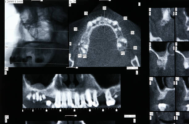
Quick Answer: Order CBCT when the clinical question involves bone — degenerative joint disease, condylar erosions, osteophytes, or trauma. Order MRI when the question involves soft tissue — disc position, disc displacement, or joint effusion. The rule I teach every referrer: bone = CBCT, disc = MRI. Some patients genuinely need both.
“Should I order CBCT or MRI for this TMJ patient?” After more than twenty years in oral and maxillofacial radiology, this remains one of the most frequent questions referring dentists send me at 3D Maxillofacial Diagnostics. And I understand why. The temporomandibular joint is the only joint most dentists are asked to image, the two modalities look at completely different tissues, and ordering the wrong one wastes the patient's money, delays diagnosis, and sometimes exposes them to radiation they did not need.
In this article, I will give you the exact decision framework I share with referring clinicians — general dentists, orofacial pain specialists, and orthodontists alike. By the end, you will be able to answer the CBCT-versus-MRI question for almost any TMJ presentation in under a minute.
Why TMJ Imaging Confuses So Many Clinicians
The confusion is not a knowledge gap — it is an anatomy problem. The temporomandibular joint is two structures in one: a bony articulation (the condyle and the glenoid fossa) and a soft tissue apparatus (the articular disc, its attachments, the retrodiscal tissue, and the joint spaces). Disease can live in either compartment, or both.
No single imaging modality shows both compartments well. CBCT is superb for mineralized tissue and nearly blind to the disc; MRI is the reference standard for the disc and inferior to CBCT for fine cortical detail. This is not a limitation of your scanner or your radiologist — it is physics. Formal diagnostic frameworks such as the DC/TMD diagnostic criteria reflect exactly this split: degenerative joint disease is confirmed on CT/CBCT, while disc displacements are confirmed on MRI.
The Core Principle: Bone = CBCT, Disc = MRI
Everything in TMJ imaging selection flows from one question: is the suspected pathology in the hard tissue or the soft tissue?
| Clinical Question | Best Modality | Why |
|---|---|---|
| Degenerative joint disease / osteoarthritis | CBCT | Erosions, osteophytes, flattening, subchondral cysts are osseous findings |
| Clicking, locking, suspected disc displacement | MRI | The disc is soft tissue — invisible on CBCT |
| Trauma, suspected condylar fracture | CBCT | High-resolution bone detail in three planes |
| Joint effusion, inflammation, marrow changes | MRI | Fluid and marrow signal require MRI sequences |
| Ankylosis, developmental anomalies (hyperplasia, hypoplasia) | CBCT | Osseous morphology and joint space assessment |
| Persistent pain with normal bone findings | MRI | Soft tissue and inflammatory causes remain |
| Suspected tumour or aggressive lesion | Both (often + medical CT) | Osseous destruction on CBCT; soft tissue extent on MRI |
The TMJ Imaging Decision Tree
Here is the step-by-step decision path I recommend to referrers. Work through it in order for any TMJ patient you are considering imaging.
STEP 1 — Is imaging indicated at all? Many TMD presentations — myofascial pain, muscle tenderness without joint signs — are managed clinically first. Image when there are joint-specific signs: crepitus, progressive limitation, occlusal change, suspected pathology, trauma, or failure of conservative therapy.
STEP 2 — Is the primary suspicion osseous? Crepitus, progressive bite change, suspected degenerative joint disease, trauma, or ankylosis → order CBCT. A dedicated TMJ protocol with both joints captured allows corrected sagittal and coronal views through each condyle.
STEP 3 — Is the primary suspicion the disc? Reciprocal clicking, intermittent locking, closed lock, or restricted opening with a soft end-feel → order MRI with both open and closed mouth views of both joints. This is the only way to classify disc displacement with or without reduction.
STEP 4 — Are there red flags? Rapidly progressive symptoms, sensory changes, swelling, or asymmetry suggesting neoplasia → order both modalities and escalate; do not wait for one result before considering the second.
STEP 5 — Still unsure? Send the clinical picture with the scan referral. A specialist can advise on modality before the patient is imaged — a two-minute question that regularly saves a wasted scan.
When CBCT Is the Right Choice for TMJ
CBCT excels at exactly one thing in the TMJ: mineralized tissue. A properly acquired TMJ CBCT lets me assess condylar morphology, cortical integrity, erosions, osteophyte formation, articular surface flattening, subchondral sclerosis and cysts, joint space dimensions, and the glenoid fossa and articular eminence. These are the findings that define degenerative joint disease, and CBCT shows them with sub-millimeter clarity at a fraction of the radiation dose of medical CT. This osseous evaluation is the core of our TMJ imaging analysis service.
Practical acquisition points for referrers: capture both joints (comparison matters diagnostically), use a field of view that includes the full condyle and fossa, and stabilize the patient well — the TMJ region is unforgiving of motion artifact.
When MRI Is the Right Choice for TMJ
MRI is the reference standard for the articular disc — its position, shape, and behaviour during opening. Reading MRI TMJ images, I evaluate disc position in the closed mouth view, whether the disc recaptures on opening (displacement with reduction) or remains displaced (without reduction), disc morphology, joint effusion, retrodiscal tissue, and marrow signal within the condyle — where changes can indicate early inflammatory or degenerative activity that bone imaging cannot yet show.
One protocol requirement I cannot emphasize enough: TMJ MRI must include both open and closed mouth views. A closed-mouth-only study can identify a displaced disc but cannot tell you whether it reduces — and that single distinction changes management. Our submission guidelines ask for both views on every TMJ MRI case for exactly this reason. Interpretation of these studies is available through our dedicated MRI TMJ interpretation service.
When Your Patient Needs Both Scans
Some presentations genuinely require both compartments assessed. The common ones in my reporting practice:
- Disc pathology (MRI) frequently coexists with secondary degenerative change (CBCT); staging both guides prognosis. Longstanding internal derangement with crepitus.
- Surgeons planning arthroscopy, arthroplasty, or joint replacement need osseous detail and disc status together. Pre-surgical TMJ workup.
- MRI detects active synovitis and effusion; CBCT documents established condylar destruction. Juvenile idiopathic arthritis and inflammatory arthropathy.
- Bony destruction patterns and soft tissue extent are both diagnostic questions. Suspected neoplasia.
When both studies exist, having one radiologist read them together is a genuine diagnostic advantage — osseous and soft tissue findings are correlated in a single narrative rather than two disconnected reports.
What About Panoramic X-rays and Other Modalities?
A word on the alternatives, because I am often asked. Panoramic radiographs can show gross condylar change and serve as an initial screen, but they distort the joint, superimpose structures, and miss early erosions — a normal panoramic does not exclude degenerative disease. Ultrasound is operator-dependent and not standard of care for the disc. Medical CT adds value mainly in trauma and tumour staging. For routine TMJ diagnosis, the working pair remains CBCT and MRI — chosen, not defaulted.
Getting the Scan Interpreted: The Step Most Practices Skip
Choosing the right modality is half the job; extracting the diagnosis from it is the other half. TMJ studies are among the most technically demanding reads in dental radiology — condylar findings hide in corrected-plane reconstructions, and disc classification on MRI takes specific training that sits at the border of dental and medical imaging. At 3D Maxillofacial Diagnostics, both study types are reported by a Canadian-certified specialist: osseous TMJ imaging analysis for CBCT volumes and dedicated interpretation of MRI TMJ images, alongside our broader CBCT interpretation services. Every report classifies findings, answers your referral question directly, and states the recommended next step.
Final Thoughts
The CBCT-versus-MRI decision for the temporomandibular joint stops being difficult the moment you anchor it to anatomy: bone equals CBCT, disc equals MRI, and complex or pre-surgical cases may need both. Work the decision tree, capture both joints, insist on open and closed mouth MRI views, and send the clinical story with the scan.
And if you would like a specialist's eyes on your next TMJ case — CBCT, MRI, or both — request a report from 3D Maxillofacial Diagnostics. One well-chosen scan, thoroughly read, is worth three ordered by default.
Frequently Asked Questions
Should I order CBCT or MRI for TMJ pain?
Start with the suspected tissue. Crepitus, bite changes, trauma, or suspected arthritis point to bone — order CBCT. Clicking, locking, or restricted opening point to the disc — order MRI with open and closed mouth views. Persistent unexplained pain with a normal CBCT is itself an indication to proceed to MRI.
What is the best imaging for TMJ disorder overall?
There is no single best. MRI is the reference standard for internal derangement and disc displacement; CBCT is the standard for degenerative joint disease and osseous pathology. The best imaging for TMJ disorder is the one matched to the clinical question — which is why the bone-versus-disc framework matters more than any default order.
Can CBCT show a displaced disc?
No. The articular disc is non-mineralized soft tissue and is essentially invisible on CBCT. Indirect signs, such as condylar positioning, are unreliable for disc diagnosis. If disc position is the question, MRI is the answer.
Does a TMJ MRI need both open and closed mouth views?
Yes. Closed mouth views establish disc position at rest; open mouth views determine whether the disc reduces during opening. The distinction between displacement with and without reduction directs management and cannot be made from a single position.
How much radiation is involved in a TMJ CBCT?
A limited field-of-view TMJ CBCT delivers a fraction of the dose of a medical CT of the same region, and MRI involves no ionizing radiation at all. Modality selection should still follow the clinical question first — the lowest-dose scan is the one that was actually indicated.